
Key Takeaways
- Advanced imaging gives surgeons a detailed view of a patient’s anatomy before robotic-assisted joint replacement surgery begins.
- Computed tomography (CT) imaging can be used to create a three-dimensional model that helps surgeons plan implant size, positioning, alignment, and bone preparation.
- During surgery, robotic systems can reference the preoperative plan and provide real-time feedback to the surgeon who is in control, promoting precision.
- Imaging and robotic technology support surgical decision-making, but they do not replace the experience, judgment, or expertise of the orthopedic surgeon in charge.
Robotic Surgery Starts with Advanced Imaging
When patients think about robotic surgery, they often picture the robotic system inside the operating room helping the medical team make cuts and placements. However, robotic systems usually begin to play a role much earlier in the treatment timeline; they are often used to upgrade imaging in surgery planning stages.
Depending on the robotic platform and procedure, computed tomography (CT) imaging may be used to create a detailed three-dimensional representation of the patient’s joint. Surgeons can then use this digital model to evaluate the individual’s unique anatomy and develop a patient-specific surgical plan before entering the operating room. Most robotic systems continually reference that plan during image-guided robotic surgery, sending helpful feedback to the surgeon to improve surgical precision.
Since 3D imaging for knee and hip replacement surgery is becoming more common throughout private medical facilities, prospective patients should educate themselves on what it entails. The guide below breaks down 3D imaging surgery and highlights its potential to help people find relief from joint pain more effectively than conventional methods.
The Evolution of Medical Imaging
Traditional X-rays were first used on humans in 1895, and they remain an important part of orthopedic evaluation because they can show joint space narrowing, bone alignment, deformity, and other changes associated with arthritis. However, X-rays are only able to provide a two-dimensional view of complex three-dimensional anatomy.
The first CT scan of a live patient happened in 1971 and raised the bar for medical imaging, as CT imaging provides additional information about the shape, orientation, and structure of the bones by showing a series of flat, two-dimensional X-ray slices at multiple angles.
CT Imaging’s Current Role in Orthopedics
CT scans are commonly used today to give surgeons a more detailed view of the anatomy they will encounter during surgery. The additional information they provide in comparison to X-rays can be particularly useful when creating arthroplasty surgical plans for people with things like:
- Significant joint deformity from arthritis
- Previous injuries that caused bone loss or structural changes
- Abnormal bone anatomy
- Previous orthopedic surgeries
Comparing X-Rays and 3D Imaging
The table below compares and contrasts X-rays with the advanced nature of CT imaging to illustrate their respective roles in joint replacement surgery.
| Feature | X-rays | CT Imaging |
| Type of image | Two-dimensional view of the joint | Gives a layered look at the joint |
| What it can show | Joint space narrowing, bone alignment, deformity, and other signs of arthritis | Detailed information about bone shape, orientation, structure, and anatomy |
| View of anatomy | Provides a general view of complex joint anatomy | Allows the surgical team to evaluate anatomy from multiple perspectives |
| Role in surgical planning | Helps evaluate the overall condition of the joint and identify certain abnormalities | Can provide additional detail for patient-specific surgical planning |
| Particularly useful for | Routine orthopedic evaluation and assessing arthritis-related changes | Patients with joint deformity, previous injuries, or unusual bone anatomy |
Even though the table above spotlights many advantages of CT imaging, one of the biggest benefits is that it can be used to create a detailed digital model for robotic-assisted surgical planning.
Turning Anatomy Into a Surgical Blueprint
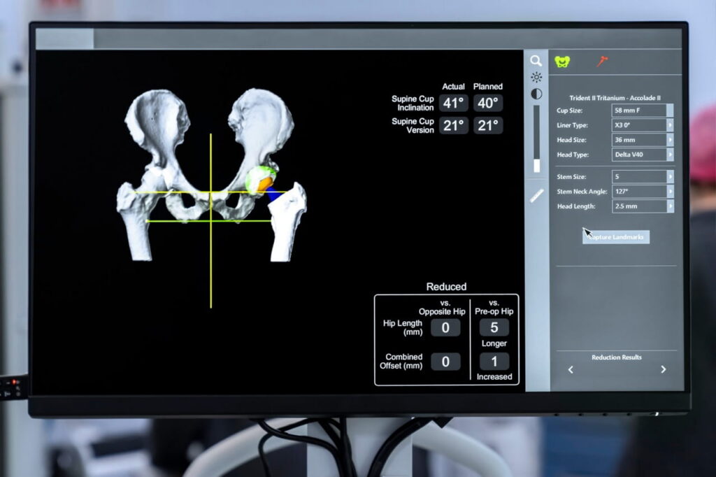
CT scan results start as a series of flat, two-dimensional X-ray slices taken from multiple angles, but a computer can stack and process these individual layers to build a complete three-dimensional digital volume of the inside of the patient’s body. This model gives the surgical team an opportunity to examine the patient’s anatomy from multiple perspectives. Rather than relying solely on measurements taken during the operation, the surgeon can evaluate important anatomical characteristics during the planning stage.
Things a Personalized Digital Model May Help Evaluate
Some things that a 3D digital model can provide extra insight into before a joint replacement surgery are:
- Bone structure – The 3D model can show the shape, size, and condition of the bones involved in the joint.
- Alignment – The digital model can illustrate the relationship between the bones and overall limb alignment.
- Implant sizing – The model can help the surgical team calculate the dimensions of the joint and available bone surface.
- Implant positioning – The 3D model often makes it easier to plan the location and orientation of implant components.
- Bone preparation – The digital model can reveal areas where the bone may need to be removed or reshaped.
- Anatomical variations – The model may highlight patient-specific abnormalities caused by arthritis, previous injuries, or prior procedures.
By working with an intricate digital representation, surgeons can build surgical plans around their patients’ unique anatomy rather than trying to conform to a generalized plan.
Planning the Procedure Before Surgery Begins
Creating a three-dimensional model is only one part of preparing for joint replacement surgery. The surgeon then uses the available information to develop an individualized surgical strategy. For a knee or hip replacement, this may include steps like:
- Determining the appropriate implant size and position
- Establishing alignment targets
- Planning the bone resections needed to prepare the joint
- Evaluating how the joint is expected to move and considering factors such as soft-tissue balance and existing deformity
By establishing important surgical parameters before entering the operating room, the team has a clearer roadmap for the procedure.
Trust Surgeons Who Prioritize Planning
The Scottsdale Center for Robotic Surgery uses advanced robotic systems to create an individualized roadmap for each patient that promotes precision.
From Digital Plan to Surgical Execution
The value of advanced imaging continues once the patient enters the operating room, as the surgical plan ironed out during early planning is usually integral for intraoperative steps as well.
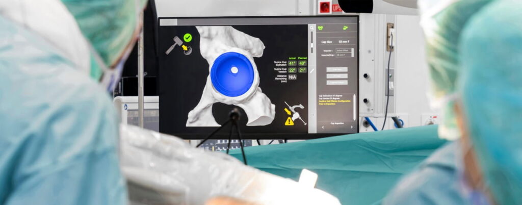
During an image-guided robotic surgery procedure, the robotic platform can reference the patient’s preoperative digital model and surgical plan. Tracking technology and intraoperative measurements help the system understand where the patient’s anatomy and surgical instruments are positioned in relation to the planned procedure, and then the system provides the corresponding feedback to the surgical team. Thanks to this process, robotic-assisted total knee arthroplasties typically offer better mechanical alignment accuracy compared to more conventional alternatives.
What Exactly Does the Robotic System Provide Feedback On?
As the procedure progresses, the surgeon can use real-time information to assess factors such as:
- Implant positioning
- Limb alignment
- Bone preparation
- Joint balance
- Range of motion
- Relationship between the actual anatomy and the planned surgical targets
If the surgical situation differs from what was anticipated during planning, the surgeon can make appropriate adjustments. This relationship between preoperative imaging, digital planning, and intraoperative guidance is a defining feature of modern robotic-assisted joint replacement.
When Advanced Imaging May Be Especially Helpful
Patient-specific imaging can be particularly valuable when the anatomy presents additional considerations for surgical planning. For example, advanced 3D imaging may help the surgical team better understand things like:
- Significant deformity
- Unusual bone geometry
- Previous surgical changes
- The extent and location of arthritic damage
This does not mean that every patient requires the same imaging approach or that advanced imaging is necessary for every joint replacement. The appropriate imaging depends on the procedure, the patient’s condition, and the surgical technology being used.
No Two Arthritic Joints Are Exactly Alike
Arthritis doesn’t affect everyone in exactly the same way. For example, one patient may have relatively uniform cartilage loss, while another may have more severe wear in one compartment.
Patient-specific imaging can help the surgeon identify anatomical characteristics that may influence the procedure before the first incision is made. A detailed three-dimensional model can provide information about bone geometry, alignment, and the extent of joint damage that a standard two-dimensional image may not fully represent.
Keep in mind, 3D imaging doesn’t automatically produce a better outcome for patients with arthritis, but the more patient-specific information the surgical team has, the better, as it can help them make informed decisions based on that individual’s unique anatomy.
Technology Doesn’t Replace Surgical Judgment
One of the most important things patients should understand about robotic-assisted surgery is that the technology does not operate independently. The robotic platform, imaging software, and planning tools function as decision-support technology. They provide information that can help the surgeon plan and execute the procedure, but they do not replace clinical judgment.
The surgeon is still responsible for things like:
- Determining whether or not joint replacement is appropriate
- Deciding which surgical approach is most suitable
- Choosing the most appropriate type of implant
- Responding to findings during the procedure
It is really important to choose the right joint replacement surgeon, as no imaging system can account for every variable encountered in an operating room—bone quality, soft-tissue tension, ligament stability, and other factors may require the surgeon to adapt the plan.
Choosing More Than a Robotic System
Your goal shouldn’t simply be to find a facility that uses 3D imaging and other advanced technology; you should focus on finding a surgical team that combines detailed information about your specific anatomy with sound surgical judgment, appropriate technology, and a comprehensive approach to care.
At the Scottsdale Center for Robotic Surgery, advanced imaging and robotic-assisted technology are incorporated into individualized surgical planning. By combining patient-specific imaging with experienced orthopedic care, the surgical team can develop a plan designed around each patient’s anatomy and treatment goals, theoretically maximizing the benefits of our outpatient surgeries.
If you’re considering joint replacement and want to understand how 3D imaging for knee or hip replacement surgery works, contact the Scottsdale Center for Robotic Surgery to schedule a consultation.